Authors: Dr. Vibha Singh*
* Associate Professor
Institution: Department of Oral and Maxillofacial Surgery, CSM Medical University, Lucknow (KGMC), India
Corresponding Author:
Dr. Vibha Singh
Associate Professor
A-43 Krishna Nagar Lucknow
Email - This email address is being protected from spambots. You need JavaScript enabled to view it.
Abstract:
Odontogenic infections are commonly the result of pericoronitis, carious teeth with pulp exposure, periodontitis, or complications of dental procedures. Periapical and panoramic x-rays are reliable initial screening instruments and are used in determining etiology. The mandibular molars are frequently one of the etiological factors. The patients with multispace infections should be hospitalized. Trismus and airway management are important considerations and may preclude the selection of other surgical approaches.
Introduction:
Dental infections are one of the most common diseases of the human body and can be a cause of death. Not until the early twentieth century, however, was a casual relationship definitely established between dental infection and severe life- threatening conditions like Ludwig's angina described over 100 years earlier.
Although the therapy has progressed, the scalpel, extraction forceps and endodontic reamer remain the keystone for the therapy for odontogenic infections as does the judicious use of antibiotics.
Prior to the antibiotic era, most serious odontogenic infections were known to be streptococcal but the problem of bacterial resistance to antibiotics soon became obvious in the oral cavity as elsewhere. The streptococcus and mixed oral flora organism remain the most commonly identified in the majority of dental infections. Considering the plethora of micro-organisms that grow luxuriantly in this wet, warm, dark and debris-strewn cavity one can only reflect with awe on the effectiveness of systemic and oral host defense mechanisms in preventing serious infection from commonplace minor trauma such as cheek biting or shedding of deciduous teeth.
There are several spaces in the head and neck region: Buccal space, Buccinator space, Parapharyngeal space, Submandibular, Sublingual, lateral pharyngeal and Pterygoid space.7 The prototype of sublingual and submandibular infection is Ludwig's angina. Ludwig's angina is caused by the extension of odontogenic infections in 70-80% of the patients.4 These conditions usually develop from an odontogenic infection especially from the 2nd and 3rd mandibular molars or as an extension of peritonsillar cellulites. Contributing factors may include teeth extraction, poor oral hygiene and trauma.
These infections are potentially life-threatening due to the spread of bacteria into the perioral facial spaces. Infection usually arises from the mandibular molars. Affected tissues are swollen and have board-like hardness. Glottis edema or spread into the mediastinum may be fatal. The principal treatment remains surgical with drainage of pus augmented with antibiotic therapy.7
Methods:
The study reviews our experience with odontogenic infections and tries to identify predisposing factors for complications.
A retrospective study population includes 100 patients with odontogenic infections from the Department of Oral and Maxillofacial Surgery C.S.M. Medical University Lucknow from 2009 to 2010. Their etiology, associated systemic diseases, bacteriology, duration of hospitalization, complications and outcomes were analyzed.
The most common age group was found to be between 40 to 60 years. The Male-Female Ratio was equal to 54:46. Only five patients had a history of diabetes mellitus. Three patients were pregnant, one in the first trimester of pregnancy and the other two were in the 3rd trimester of pregnancy. Out of 100 patients, 94 patients underwent extraction of the offending teeth with surgical incision and drainage. In six patients, there was post-extraction space infection. They all were referred by dentists.
Pus culture was sterile in 90% of the patients because patients were referred by dental surgeons who were treating them with broad spectrum antibiotics.. 6% of the patients showed gram positive cocci .
Empirically, IV antibiotic therapy for odontogenic infection is commonly chosen. In most of the patients, third generation cephalosporins along with metronidazole was advised. In other patients, Amoxycillin with clavulanic acid with metronidazole were used. The patients were also hydrated and medical treatment was given for associated conditions.
Investigations: An orthopantomogram was taken to identify offending teeth. In a few cases of temporal space infection, CT scan was used to evaluate the extent of infection.4
Six patients developed infection after extraction and 94 patients were having odontogenic foci of infection. The most commonly involved teeth were found to be the mandibular first molar, followed by the 3rd molar. In seven cases, the maxillary molar was involved.
The most commonly involved space was the submandibular space (42%), followed by buccal (28%) and masseteric space (15%). In six patients, there was bilateral involvement of the submandibular and submental spaces diagnosed as Ludwig's angina.
In five patients, the temporal space was involved and severe pain and trismus was the main complaint of the majority of the patients, which is the most common feature in odontogenic infections.
Table I: Severity Score-- Odontogenic Infections
- Severity Score 1: Low risk to airway or vital structure. Anatomic Site: Vestibular, Subperiosteal, Space of the body of the mandible, Infraorbital, Buccal
- Severity Score 2: Moderate risk to airway or vital structure. Anatomic Site: Submandibular, Submental, Sublingual, Pterigomandibular, Submasseteric, Superficial temporal, Deep temporal
- Severity Score 3: High risk to airway or vital structure. Anatomic Site: Retropharyngeal
- Severity Score 4: Extreme high risk to airway or vital structure. Anatomic Site: Danger space 4, Mediastinum, Intracranial infection
Results:
Table 2 shows a breakdown of our patient population and the extent of the disease. There was not a sex prominence. The peak age of the patient was 40 to 60 years. The mandibular teeth were more commonly involved than the maxillary teeth, 3.7 to one. Molars and PreMolars were the most common etiology of the infection. No abscesses were caused by the Canine or Incisors. Five patients were diabetics and three were pregnant.
Six patients were treated with incision and drainage. 94 were treated with extraction, incision and drainage. No patient required a tracheotomy. No patient died and all were cured of the infection. Cultures were only positive in six patients (negative in 90) which grew gram positive cocci.

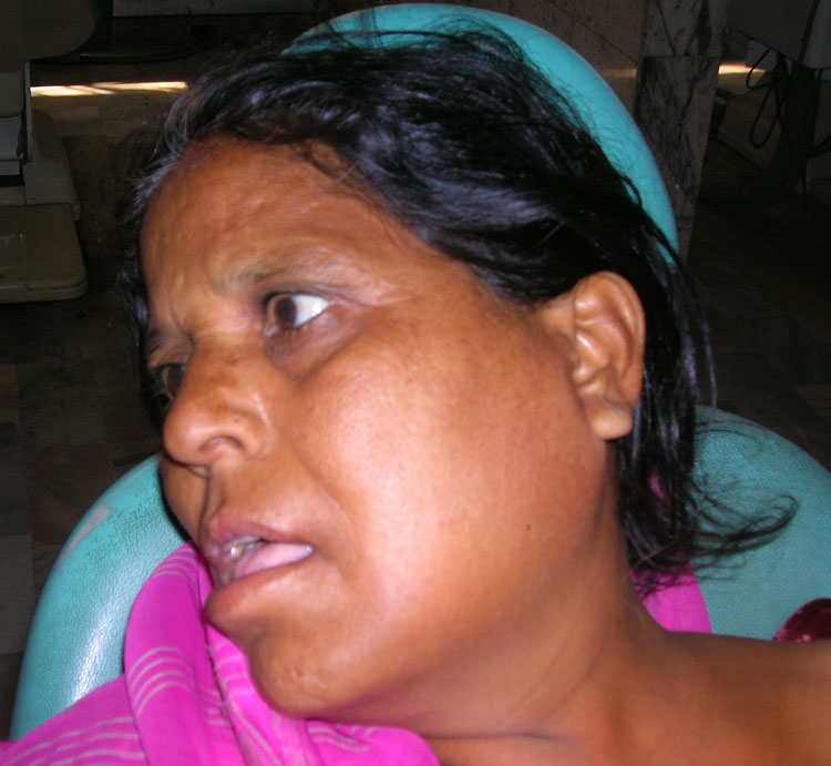
Patient on the right is a 40 year old female with a submandibular and buccal space infection.

35 year old male with an infection involving the buccal and temporal space.

Axial CT scan showing involvement of the temporal space with infection.

Coronal CT scan showing involvement of the buccal and temporal space with infection.
Table 2: Patient Population and Extent of Disease.
| Parameter | Variable | Number |
| Sex | Male Female |
54 46 |
| Age | 0-20 yrs 20-40 yrs 40-60 yrs 60 and above |
17 33 44 16 |
| Teeth | Mandibular Maxillary |
63 17 |
| Spaces Involved | Buccal Submandibular Submental Temporal Lateral Pharyngeal Ludwig's Space |
20 15 42 5 0 6 |
| Symptoms | Swelling Trismus Dysphagia Dyspnea |
100 85 6 2 |
Discussion:
Odontogenic infections generally pass through three stages before they resolve during the first 1 to 3 days. The swelling is soft, mildly tender and doughy in consistency. Between two to five days, the swelling becomes hard, red and exquisitely tender. Its borders are diffuse and spreading. Between the fifth and seventh days, the center of the cellulites begins to soften and the underlying abscess undermines the skin and mucosa and makes it compressible and shiny. At this stage, the infection is fluctuant. The final stage of odontogenic infection is resolution which generally occurs after spontaneous or surgical drainage of an abscess cavity. Trismus is an ominous sign in the patient with suspected odontogenic infection.5
Odontogenic infections rarely extend beyond the jawbone barrier into the deep space of the face and neck but once they occur they are often difficult to assess accurately by clinical and conventional radiographs and the outcome may be potentially life threatening. CT scan has been used to assess and evaluate deep neck infections of odontogenic origin. It is useful to depict the extent of infection and to plan treatment of extensive odontogenic infection.4
Maxillary space infection can originate from either the maxillary or mandibular teeth. Temporal space can be divided into superficial and deep temporal space. The superficial temporal space extends superiorly to the pericranium, lateral to the temporalis muscle and medial to the temporoparital fascia. Inferiorly, this space is continuous with the masseteric space. The deep temporal space extends superiorly to the attachment of the temporalis muscle to the inferior temporal crest, lateral to the temporal bone and deep to the temporalis muscle. Inferiorly, this space is continuous with the infratemporal space. The temporal space along with the infratemporal, masseteric and pterigo mandibular space can be grouped together as masticator space.
The masticator space is defined by the superficial layer of the deep cervical fascia as it splits at the inferior border of the mandible. The lateral portion covers the masseter as it connects to the zygomatic arch and continues on to cover the temporalis muscle. The medial portion follows the medial pterygoid, superiorly, then continues with the levator veli palatine muscle. The medial portion follows the medial pterygoid, superiorly, then continues with the levator palatine fascia to skull base. Space adjacent to the masticator space are the parotid space, posteriorly, parapharyngeal space, medially, and submandibular and sublingual space, inferiorly.1
Necrotizing fasciitis is occasionally found in the head and neck and is frequently due to odontogenic sources. It is a rapidly spreading infection that follows the platysma muscle down the neck and to the anterior chest wall. Diabetes and alcoholism have been shown to be significant predisposing factors, whereas, high-risk medical conditions, delay in surgery and mediastinitis are associated with increased mortality.
A suspicion of necrotizing fasciitis is a surgical emergency ,requiring broad spectrum antibiotics, repeated surgical dressing, drainage, intensive medical supportive care including fluid calcium and possibly blood transfusion.5
Conclusion:
The diagnosis of odontogenic infections is usually obvious. If not, a radiograph and a CT scan is performed. Treatment of odontogenic infection is maintenance of airway patency, surgical incision, extraction of offending teeth, drainage and antibiotics.
References:
1. Morrison A, Brady J. Temporal space infection secondary to mandibular extraction. Oral Health Journal. June 2009.
View Article
2. Bratton TA, Jackson DC, Nkungula-Howlett T, Williams CW, Bennett CR. Management of complex multi-space odontogenic infections. J Tenn Dent Assoc. 2002 Fall;82(3):39-47. View Abstract
3. Huang TT, Liu TC, Chen PR, Tseng FY, Yeh TH, Chen YS. Deep neck infection: analysis of 185 cases. Head Neck. 2004 Oct;26(10):854-60. View Abstract
4. Yonetsu K, Izumi M, Nakamura T. Deep facial infections of odontogenic origin: CT assessment of pathways of space involvement. AJNR Am J Neuroradiol. 1998 Jan;19(1):123-8. View Abstract
5. Neal D. Futran MD, DMD Peterson's Principles of Oral and Maxillo Facial Surgery. 2nd edition by Michael Miloro, B.C. Decker, Inc., Hamilton, 2004, 1500 pp View Book
6. Ariji Y, Gotoh M, Kimura Y, Naitoh M, Kurita K, Natsume N, Ariji E. Odontogenic infection pathway to the submandibular space: imaging assessment. Int J Oral Maxillofac Surg. 2002 Apr;31(2):165-9. View Abstract
7. Cervico-facial infection and Ludwigs angina. exodontias .info March 11, 2011. View Article